|
"Few students of biological science are trained to look beyond biochemistry and pathobiology to apply two principles fundamental to the architect and mechanical engineer; these are sound structure and sufficient bracing. The application of these principles seems essential to the investigation of hip dysplasia."
- Wayne H. Riser, DVM; Founder and first Program Director of the Orthopedic Foundation for Animals |
|
Let's start with a point made in a previous blog (The 10 most important things to know about canine hip dysplasia), that puppies are born with "perfect", normal hips. Of course, they're puppy hips and not adult hips, but they are quite remarkable.
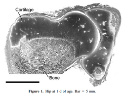
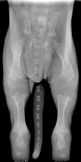
A newborn puppy looks like it has no joints at all on an x-ray. This is because the ends of the long bones and many parts of the pelvis are soft cartilage at birth. Because cartilage doesn't show up on x-rays, a radiograph of a newborn puppy can look like an apparition of a spooky, disarticulated body. But this is just nature's way of providing enough support to be able to move around while the skeletal grows rapidly in the first few months of life.
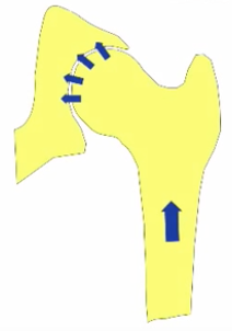
The hip joints are also formed of cartilage at birth and are little more than a round ball at the end of the femur that sits in a depression in the pelvis where the hip socket will be.
|
This seems truly magical, but there's a catch. If, for some reason, the head of the femur is not tightly held in the hip socket, development will go awry. What results is "developmental hip dysplasia"; a malformed hip socket. In dogs, this is canine hip dysplasia.
Wayne Riser, who studied hip dysplasia in dogs for many years and was also the founder and first director of the Orthopedic Foundation for Animals, explained it this way (1975):
|
"In all mammalian embryos, the hip is laid down as a single unit from mesenchymal tissue, and it develops normally as long as the components are left in full congruity. The hip is normal at some time in the development of the mammal, and abnormal development occurs only when stresses pull the components apart.
In the dog, the hip is normal at birth. Intrauterine stresses are not sufficient to produce incongruity of the hip. The first time such forces are great enough is when the pup begins to take its position to nurse. Observations of the disease in man, dog, and a number of other mammals for many years have culminated in the conviction that the bony changes of hip dysplasia, regardless of species, occur because the soft tissues do not have sufficient strength to maintain congruity between the articular surfaces of the femoral head and the acetabulum." |
It might be hard to visualize what's going on, so here's a great video that shows how this can happen. It is about hip dysplasia in humans, but the anatomy is the same and the manual diagnostic techniques (Barlow and Ortolani tests) they demonstrate are used for dogs as well. The last 20 minutes or so are about examination of the human infant so you might want to skip that, and for some reason it's a 30 minute video that repeats, so it's not really an hour long.
(Check out the shifty eyes on that baby. I think this might be the very same infant that went on to become Ally McBeal's famous "dancing baby" hallucination. If you missed that somehow, you can catch it here.)
As Riser described, looseness or laxity of the hip joint is the prerequisite for the development of hip dysplasia. If the head of the femur is not positioned properly in the hip socket during development, the mechanical forces that stimulate bone deposition in the joint will be abnormal and the result will be a dysplastic hip.
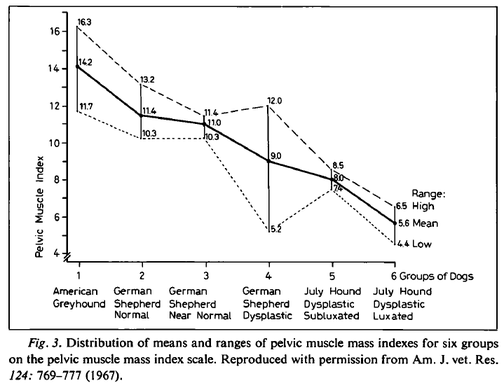
In breeds like the Greyhound, which very rarely suffer from hip dysplasia, the muscles that support the hip are exceptionally well developed. Riser found that less pelvic muscle mass was associated with higher risk of dysplasia. This even held within a breed: in both German Shepherds and July Hounds, dogs with a higher pelvic mass index had better hips on average.
|
|
|
Unfortunately, most breeds of dogs don't have the exceptional pelvic muscles of the Greyhound at birth, which are the result of selection over hundreds of generations to produce a dog with exceptional speed. The Bernese Mountain Dog, on the other hand, was bred to have the size, strength, and substance necessary for drafting (cart hauling). In fact, anatomical differences among breeds are reflected in propensity to develop hip dysplasia.
After studying radiographs of tens of thousands of dogs in dozens of breeds, Riser (1975) found that a pattern began to emerge.
|
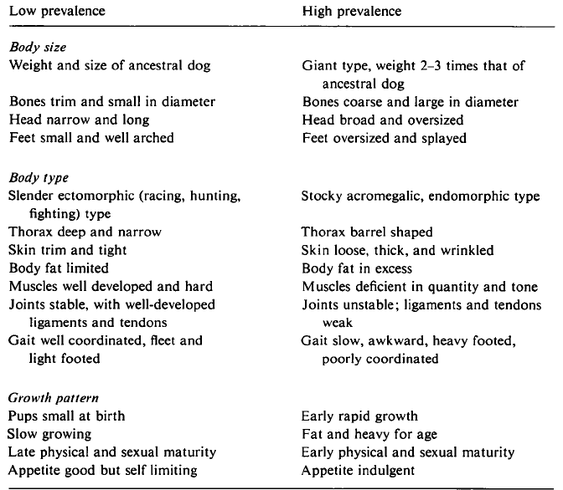
There was...a strong correlation between body form, size, growth rate, quantity of subcutaneous fat, type of connective tissue, pelvic muscle mass, and the general body type of the different breeds and the prevalence of hip dysplasia. Recently, we have identified certain general characteristics of a breed that increase the risk of hip dysplasia.
Body Size The breeds with the lowest percentage of hip dysplasia were near the size of the ancestral dog. The bones were small in diameter and smooth, the feet were small and well arched, and the shape of the head was long and narrow. The giant breeds with a high percentage of hip dysplasia were two to three times larger than the ancestral dog. Their bones were coarse and large in diameter, with prominent protrusions and depressions. The feet were large and splayed, and the head was wide and oversized. Body Type In general, the body conformation of the breeds with the lowest percentage of hip dysplasia was slender and trim. The skin was thin, smooth, and stretched tightly over the underlying tissues. The muscles were prominent, hard, and full-bellied. At dissection in these breeds, the skin and subcutaneous tissues and fascia rarely contained over 1-2% fat by weight. The joint ligaments were well developed; the fibers were coarse, closely packed, and relatively free of fat. The well-formed pelvic and thigh muscles were attached to broad, coarse tendons that were securely attached to the bones. This group of dogs is fleet-footed and well-coordinated in their movements. Of the high-risk group, the four breeds of the giant type were not only two to three times the size of the ancestral dog, but their body conformation was heavy, round, and stocky. Acromegalic characteristics were present to some extent in all four breeds. The skin was thicker than that in the other group; it lay in folds over the head and neck. At dissection, the thickened skin was infiltrated with fat. Fat was also abundant in the subcutaneous and fascia spaces and commonly accounted for 5-10% of the weight of the soft tissues of the hindquarters. In comparison with the other group, the muscles were less prominent and less developed. Fat also was infiltrated into the tendons and ligaments. The fibers of these two structures were smaller in diameter than those of the low-risk group. The gait of the giant breeds was less graceful and slower than that of the smaller breeds. Growth Pattern The group of breeds with the highest percentage of hip dysplasia grew and matured more rapidly than did those in the low-risk group. We have observed this in several studies. Starting at birth, this group gained rapidly. The pups of these breeds were aggressive eaters, both as they nursed and as they began to take supplemental food. The 38 breeds, when ranked according to the highest prevalence for hip dysplasia, with few exceptions exhibited a gradual shift from the poorly muscled and poorly coordinated, acromegalic type giants at the top, to the lowest percentage of hip dysplasia at the bottom, characterized by the breeds that were sleek, tight skinned, highly coordinated and well muscled. These correlations and observations support previous findings that the poorly muscled and coordinated breeds have a high percentage of hip dysplasia, whereas the well-muscled and highly coordinated types are relatively free of the disease. Selection for acceleration in growth created dogs with excessive fat and weight at an early age. This has resulted in lowered dynamic and biomechanical efficiency of the hip joint. The young dog that carries excessive weight runs the risk of over-extending the supporting soft tissues, and injury to these tissues results in pulling apart (subluxation) of the joint components. This results in changes that have been recognized as hip dysplasia. This is not a new concept. It was pointed out as long ago as 3 centuries that 'Muscles and bone are inseparably associated and connected, between muscle and bone there can be no change in one but it is correlated with changes within the other'. |
Riser summarized these findings in a table:
The factors that cause hip laxity and the development of dysplasia occur in the first few months of the puppy's life. Sometime between birth, when the cartilagenous tissues of the hip joint have the capacity to become a perfectly constructed ball and socket joint, and when the puppy is four or five months old, the soft tissues of the pelvis fail to provide the support necessary to keep the femoral head properly seated in the hip socket. As a result, the joint doesn't develop properly, then abnormal biomechanical forces exacerbated by environmental factors such as weight and inappropriate exercise begin the cycle of damage and inflammation that result in dysplasia and osteoarthritis.
|
In very young human and canine subjects with unstable coxofemoral joints, hip dysplasia can be prevented and the instability corrected if the congruity of the components of the hip joint is maintained and if femoral subluxation does not occur. If proper congruity cannot be maintained, the hip joint becomes malformed in a relatively short time. The changes in the bones and cartilage of the hip are thus the indirect result of failure of the soft tissues to support full congruity of the bony components of the hip.
Few genes so far analyzed directly affect osseous structures. [This is still true.] The shape of bones reflects changes by biomechanical stresses. The spread of hip dysplasia centers around the genetic transmission and heritability of certain body size, type, conformation, movement, growth pattern, and temperament. This conclusion is based on the facts that the prevalence of hip dysplasia is approximately the same in a number of breeds with similar body characteristics and there is no gene flow between these purebred breeds. Since these facts must be respected, biomechanical and environmental factors associated with certain body conformation and size must be considered as the causes. |
According to Riser, the risk factors for hip dysplasia are related to the features of a dog and its breed that result in a mismatch between the forces necessary to maintain congruency of the hip joint and the support the soft tissues are able to provide during the critical first few months of life. By the time the puppy is 6 months old, the strength of the supporting tissues and ossification of the bones should be adequate to prevent the development of hip dysplasia under normal circumstances. Prevention of hip dysplasia will come both from reducing the genetic risk factors through selection and from avoiding situations that could result in joint instability or incongruity.
|
The literature review and report of our work presented here have revealed a basis for optimism in controlling and reducing the prevalence of hip dysplasia in both man and animals...In children, it is basically the test for hip laxity at birth, and for the dog it is restriction of breeding only those animals with radiographically normal hips. Both in children and dogs, hip dysplasia is, for the most part, a ‘man-made’ problem and can be controlled if man will use the tools at his disposal.
These findings uphold the validity of our two premises that (1) hip dysplasia occurs only if hip joint instability and joint incongruity are present in the young child or animal; and (2) the disease can be prevented if hip joint congruity can be maintained until ossification makes the acetabulum less plastic and the abductor muscles and supporting soft tissues become sufficiently strong and functional to prevent femoral head subluxation. |
Genetic selection and management of environmental risks are both necessary to lower the frequency of hip dysplasia. For selection to be effective, we need to recognize that some of the traits that are valued as part of breed type are themselves risk factors for development of hip dysplasia. Selecting for faster growth and ever increasing size will confound efforts to produce dogs with better hips. Likewise, inadequate weight management of both puppies and adults is an environmental factor that dramatically increases the risk of developing dysplastic hips. Finally, we should use a little common sense about activities that are appropriate for puppies (see below...).
Understanding Hip and Elbow Dysplasia
Starts 4 January 2016
Wayne Riser DVM was on the faculty of the vet school at the Univ. of Pennsylvania and was also the founder and first Program Director of OFA. In 1975, he published a collection of papers in the journal Veterinary Pathology that,taken together, represented a summary of what was known at the time about the disease of hip dysplasia in the dog. Much of what Riser had to say about anatomy, growth, development, and biomechanics forty years ago is just as relevant to breeders and dog owners today.
Understanding Hip & Elbow Dysplasia
Next class starts
2 May 2022
Sign up now!
***************************************
Visit our Facebook Groups
ICB Institute of Canine Biology
...the latest canine news and research
ICB Breeding for the Future
...the science of dog breeding
