The Institute of Canine Biology
  • HOME
  • Blog
  • Courses
    • COI BootCamp (FREE!)
    • Basic Population Genetics (FREE)
    • The Science of Canine Husbandry
    • Managing Genetics For the Future >
      • Syllabus - Managing Genetics for the Future
    • The Biology of Dogs (Open Reg )
    • DNA For Dog Breeders >
      • Syllabus - DNA for Dog Breeders
      • Open Reg - DNA For Dog Breeders
    • Understanding Hip & Elbow Dysplasia >
      • Open Reg - Understanding Hip & Elbow Dysplasia
    • Genetics of Behavior & Performance >
      • Syllabus - Genetics Behavior & Performance
      • Open Reg - Genetics of Behavior & Performance (Open Reg)
    • Strategies for Preservation Breeding >
      • Open Reg - Strategies for Preservation Breeding
    • Group Discounts
    • MORE FREE COURSES >
      • Quickie Genetics (Free!)
      • Heredity & Genetics (Free!)
      • Useful Genetics (Free!)
      • Basic Genetics Videos
  • Breed Preservation
    • Breed Status
    • Breeding for the future >
      • BFF Breed Groups
    • The "Elevator Pitch"
    • What's in the Gene Pool?
    • The Pox of Popular Sires
    • What population genetics can tell us about a breed
    • What population genetics can tell you...Tollers & Heelers
    • How to use kinship data
    • Using EBVs to breed better dogs >
      • How population size affects inbreeding
      • EBV Examples
    • How to read a dendrogram
    • Global Pedigree Project >
      • The Database
    • Finding the genes without DNA
    • How to read a heat map
  • Health Data
    • Bloat (Purdue Study)
    • Body Condition Score >
      • % Dysplastic vs BCS
    • Breed Comparions
    • Cancer
    • Cardiac
    • Cataracts
    • Caesareans
    • Deafness
    • Degenerative Myelopathy
    • Elbow Dysplasia
    • Epilepsy
    • Genetic Diversity
    • Genetic Diversity (MyDogDNA)
    • Hip Dysplasia >
      • Hip Dysplasia (Hou et al 2013)
    • Inbreeding Effects
    • Inbreeding (Gubbels)
    • Inbreeding (Dreger)
    • Lifespan
    • Litter size
    • Metabolic
    • mtDNA
    • Orthopedic
    • Mode of Inheritance
    • Patella Luxation
    • Thyroid
    • Portosystemic shunt
    • Purebred vs Mixed (UC Davis)
    • Purebred vs Mixed Breed (Bonnett)
    • Spay & Neuter Effects
    • Calboli et al 2008
    • Hodgman (1963)
    • Scott & Fuller (1965)
    • Stockard: Purebred crosses
    • Summers (2011)
  • Projects
    • How To Interpret Breed Analyses
    • Afghan Hound
    • More details about the Toller study
    • Belgian Tervuren >
      • Belgian Terv p2
      • Belgians- why population size matters
    • Bernese Mountain Dog
    • Boxer
    • Brussels Griffon
    • Bullmastiff
    • Canaan Dog >
      • Canaan analyses
    • Cesky Terrier >
      • Cesky genetic history
    • Chinook
    • Curly-coated Retriever
    • Doberman
    • Entelbucher Mountain Dog
    • Flatcoat Retriever
    • French Bulldog
    • German Shorthair
    • Golden Retriever >
      • Golden Retriever Pedigree Charts
    • Irish Water Spaniel >
      • IWS (6 Nov 17)
    • Labrador Retriever
    • Manchester Terrier
    • Mongolian Bankhar >
      • Research Updates
      • Bankhar 1
    • Norwegian Lundehund
    • Plummer Terrier
    • Otterhound
    • Portuguese Water Dog >
      • Portuguese Water Dog (pt 2)
    • Ridgeback
    • Schipperke
    • Standard Poodle >
      • The Problem With Poodles
      • 3poodle pedigree charts
      • 3Poodle Wycliff dogs
      • Poodle Genetics
    • Tibetan Spaniel
    • Tibetan Mastiff
    • West Highland White Terrier
    • Whippet
    • Wirehaired Pointing Griffons
    • UK KC Graphs >
      • UK KC Breed Status
      • UK Groups
      • KC Gundogs
      • KC Hounds
      • KC Terriers >
        • Terriers (select breeds)
      • KC Pastoral
      • KC Toys
      • KC Working
      • KC Utility
      • Australian KC
    • Breed outcrossing programs
  • Resources
    • Genetics Databases
    • Stud Books >
      • American Kennel Club stud books
      • Field Dog stud books
      • The Kennel Club (UK)
    • Learn
    • Videos about dog genetics
    • The Amazing Things Dogs Do! (videos) >
      • Livestock Management
      • Livestock guarding
      • Transportation, exploration, racing
      • Conservation & wildlife management
      • Detection Dogs
      • Medicine & Research
      • Entertainment
      • AKC/CHF Podcasts
    • Read & Watch
    • Bookshelf
  • Preventing Uterine Inertia

How do hips become dysplastic?

12/23/2015

 
By Carol Beuchat PhD
"Few students of biological science are trained to look beyond biochemistry and pathobiology to apply two principles fundamental to the architect and mechanical engineer; these are sound structure and sufficient bracing. The application of these principles seems essential to the investigation of hip dysplasia."
 - Wayne H. Riser, DVM; Founder and first Program Director of the Orthopedic Foundation for Animals

Let's start with a point made in a previous blog (The 10 most important things to know about canine hip dysplasia), that puppies are born with "perfect", normal hips. Of course, they're puppy hips and not adult hips, but they are quite remarkable.  

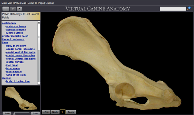
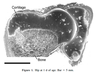
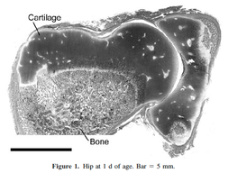
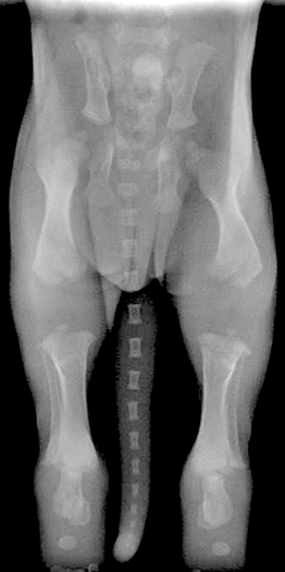
A newborn puppy looks like it has no joints at all on an x-ray. This is because the ends of the long bones and many parts of the pelvis are soft cartilage at birth. Because cartilage doesn't show up on x-rays, a radiograph of a newborn puppy can look like an apparition of a spooky, disarticulated body. But this is just nature's way of providing enough support to be able to move around while the skeletal grows rapidly in the first few months of life.
The hip joints are also formed of cartilage at birth and are little more than a round ball at the end of the femur that sits in a depression in the pelvis where the hip socket will be.
Picture
Picture
As the puppy grows, the formation of the bony structures that will become the hip joint is not programmed by genes. Instead, the forces on the joint stimulate the deposition of bone in the right places to form an articulating ball and socket joint. As long as the head of the femur stays seated where it belongs in the developing hip socket, the hip joint should form perfectly.

This seems truly magical, but there's a catch. If, for some reason, the head of the femur is not tightly held in the hip socket, development will go awry. What results is "developmental hip dysplasia"; a malformed hip socket. In dogs, this is canine hip dysplasia.

​Wayne Riser, who studied hip dysplasia in dogs for many years and was also the founder and first director of the Orthopedic Foundation for Animals, explained it this way (1975):
"In all mammalian embryos, the hip is laid down as a single unit from mesenchymal tissue, and it develops normally as long as the components are left in full congruity. The hip is normal at some time in the development of the mammal, and abnormal development occurs only when stresses pull the components apart.

In the dog, the hip is normal at birth. Intrauterine stresses are not sufficient to produce incongruity of the hip. The first time such forces are great enough is when the pup begins to take its position to nurse.

​Observations of the disease in man, dog, and a number of other mammals for many years have culminated in the conviction that the bony changes of hip dysplasia, regardless of species, occur because the soft tissues do not have sufficient strength to maintain congruity between the articular surfaces of the femoral head and the acetabulum."
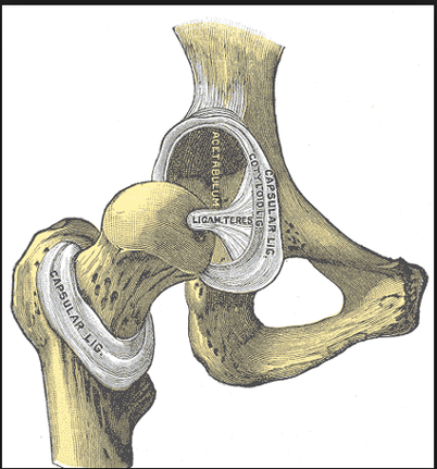
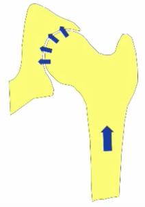
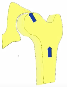
Riser is saying here that muscles, ligaments, and tendons ("soft tissues") normally keep the head of the femur properly seated in the developing socket. But if there are abnormal forces on the joint, the soft tissues might be inadequate to stabilize the joint, and malformation of the socket - hip dysplasia - will occur.

It might be hard to visualize what's going on, so here's a great video that shows how this can happen. It is about hip dysplasia in humans, but the anatomy is the same and the manual diagnostic techniques (Barlow and Ortolani tests) they demonstrate are used for dogs as well. The last 20 minutes or so are about examination of the human infant so you might want to skip that, and for some reason it's a 30 minute video that repeats, so it's not really an hour long.

(Check out the shifty eyes on that baby. I think this might be the very same infant that went on to become Ally McBeal's famous "dancing baby" hallucination. If you missed that somehow, you can catch it here.)
​
As Riser described, looseness or laxity of the hip joint is the prerequisite for the development of hip dysplasia. If the head of the femur is not positioned properly in the hip socket during development, the mechanical forces that stimulate bone deposition in the joint will be abnormal and the result will be a dysplastic hip.

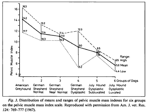
In breeds like the Greyhound, which very rarely suffer from hip dysplasia, the muscles that support the hip are exceptionally well developed. Riser found that less pelvic muscle mass was associated with higher risk of dysplasia. This even held within a breed: in both German Shepherds and July Hounds, dogs with a higher pelvic mass index had better hips on average.
Picture
Riser 1975
The exceptional hip stability provided by the pelvic muscles of Greyhounds is evident even in newborns. Check out the one day old Greyhound puppies, standing and climbing to nurse, and looking like Ninjas compared to the desperately cute but nearly helpless baby Berners.

Unfortunately, most breeds of dogs don't have the exceptional pelvic muscles of the Greyhound at birth, which are the result of selection over hundreds of generations to produce a dog with exceptional speed. The Bernese Mountain Dog, on the other hand, was bred to have the size, strength, and substance necessary for drafting (cart hauling). In fact, anatomical differences among breeds are reflected in propensity to develop hip dysplasia.

After studying radiographs of tens of thousands of dogs in dozens of breeds, Riser (1975) found that a pattern began to emerge.
There was...a strong correlation between body form, size, growth rate, quantity of subcutaneous fat, type of connective tissue, pelvic muscle mass, and the general body type of the different breeds and the prevalence of hip dysplasia. Recently, we have identified certain general characteristics of a breed that increase the risk of hip dysplasia.

Body Size
The breeds with the lowest percentage of hip dysplasia were near the size of the ancestral dog. The bones were small in diameter and smooth, the feet were small and well arched, and the shape of the head was long and narrow. The giant breeds with a high percentage of hip dysplasia were two to three times larger than the ancestral dog. Their bones were coarse and large in diameter, with prominent protrusions and depressions. The feet were large and splayed, and the head was wide and oversized.

Body Type​
In general, the body conformation of the breeds with the lowest percentage of hip dysplasia was slender and trim. The skin was thin, smooth, and stretched tightly over the underlying tissues. The muscles were prominent, hard, and full-bellied. At dissection in these breeds, the skin and subcutaneous tissues and fascia rarely contained over 1-2% fat by weight. The joint ligaments were well developed; the fibers were coarse, closely packed, and relatively free of fat. The well-formed pelvic and thigh muscles were attached to broad, coarse tendons that were securely attached to the bones. This group of dogs is fleet-footed and well-coordinated in their movements. Of the high-risk group, the four breeds of the giant type were not only two to three times the size of the ancestral dog, but their body conformation was heavy, round, and stocky. Acromegalic characteristics were present to some extent in all four breeds. The skin was thicker than that in the other group; it lay in folds over the head and neck. At dissection, the thickened skin was infiltrated with fat. Fat was also abundant in the subcutaneous and fascia spaces and commonly accounted for 5-10% of the weight of the soft tissues of the hindquarters. In comparison with the other group, the muscles were less prominent and less developed. Fat also was infiltrated into the tendons and ligaments. The fibers of these two structures were smaller in diameter than those of the low-risk group. The gait of the giant breeds was less graceful and slower than that of the smaller breeds.

Growth Pattern
The group of breeds with the highest percentage of hip dysplasia grew and matured more rapidly than did those in the low-risk group. We have observed this in several studies. Starting at birth, this group gained rapidly. The pups of these breeds were aggressive eaters, both as they nursed and as they began to take supplemental food.

The 38 breeds, when ranked according to the highest prevalence for hip dysplasia, with few exceptions exhibited a gradual shift from the poorly muscled and poorly coordinated, acromegalic type giants at the top, to the lowest percentage of hip dysplasia at the bottom, characterized by the breeds that were sleek, tight skinned, highly coordinated and well muscled. These correlations and observations support previous findings that the poorly muscled and coordinated breeds have a high percentage of hip dysplasia, whereas the well-muscled and highly coordinated types are relatively free of the disease.

Selection for acceleration in growth created dogs with excessive fat and weight at an early age. This has resulted in lowered dynamic and biomechanical efficiency of the hip joint. The young dog that carries excessive weight runs the risk of over-extending the supporting soft tissues, and injury to these tissues results in pulling apart (subluxation) of the joint components. This results in changes that have been recognized as hip dysplasia. This is not a new concept. It was pointed out as long ago as 3 centuries that 'Muscles and bone are inseparably associated and connected, between muscle and bone there can be no change in one but it is correlated with changes within the other'.

Riser summarized these findings in a table:
Picture
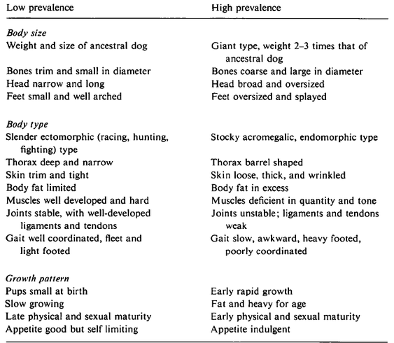
You will recognize that many of these traits are features of breed type, and of course most are under direct genetic selection or are secondary to some other trait under selection. As it seems to go in the competitive show world, many of the traits on the "High Prevalence" list have become progressively more exaggerated through artificial selection - the large size gets larger, heavy bone gets heavier, a broader head gets broader. And to make matters worse, the desire for dogs that "finish" (become champions) at ever younger ages drives selection for rapid growth, which overloads the immature skeleton and muscle systems. It seems that we are trying to select against hip dysplasia at the same time as we select for many traits that increase the risk.

The factors that cause hip laxity and the development of dysplasia occur in the first few months of the puppy's life. Sometime between birth, when the cartilagenous tissues of the hip joint have the capacity to become a perfectly constructed ball and socket joint, and when the puppy is four or five months old, the soft tissues of the pelvis fail to provide the support necessary to keep the femoral head properly seated in the hip socket. As a result, the joint doesn't develop properly, then abnormal biomechanical forces exacerbated by environmental factors such as weight and inappropriate exercise begin the cycle of damage and inflammation that result in dysplasia and osteoarthritis. 
Wayne Riser showed 40 years ago that ​
In very young human and canine subjects with unstable coxofemoral joints, hip dysplasia can be prevented and the instability corrected if the congruity of the components of the hip joint is maintained and if femoral subluxation does not occur. If proper congruity cannot be maintained, the hip joint becomes malformed in a relatively short time. The changes in the bones and cartilage of the hip are thus the indirect result of failure of the soft tissues to support full congruity of the bony components of the hip.

Few genes so far analyzed directly affect osseous structures.
[This is still true.] The shape of bones reflects changes by biomechanical stresses.

The spread of hip dysplasia centers around the genetic transmission and heritability of certain body size, type, conformation, movement, growth pattern, and temperament. This conclusion is based on the facts that the prevalence of hip dysplasia is approximately the same in a number of breeds with similar body characteristics and there is no gene flow between these purebred breeds. Since these facts must be respected, biomechanical and environmental factors associated with certain body conformation and size must be considered as the causes.

According to Riser, the risk factors for hip dysplasia are related to the features of a dog and its breed that result in a mismatch between the forces necessary to maintain congruency of the hip joint and the support the soft tissues are able to provide during the critical first few months of life. By the time the puppy is 6 months old, the strength of the supporting tissues and ossification of the bones should be adequate to prevent the development of hip dysplasia under normal circumstances. Prevention of hip dysplasia will come both from reducing the genetic risk factors through selection and from avoiding situations that could result in joint instability or incongruity.
The literature review and report of our work presented here have revealed a basis for optimism in controlling and reducing the prevalence of hip dysplasia in both man and animals...In children, it is basically the test for hip laxity at birth, and for the dog it is restriction of breeding only those animals with radiographically normal hips.  Both in children and dogs, hip dysplasia is, for the most part, a ‘man-made’ problem and can be controlled if man will use the tools at his disposal.

These findings uphold the validity of our two premises that (1) hip dysplasia occurs only if hip joint instability and joint incongruity are present in the young child or animal; and (2) the disease can be prevented if hip joint congruity can be maintained until ossification makes the acetabulum less plastic and the abductor muscles and supporting soft tissues become sufficiently strong and functional to prevent femoral head subluxation.

Genetic selection and management of environmental risks are both necessary to lower the frequency of hip dysplasia. For selection to be effective, we need to recognize that some of the traits that are valued as part of breed type are themselves risk factors for development of hip dysplasia. Selecting for faster growth and ever increasing size will confound efforts to produce dogs with better hips. Likewise, inadequate weight management of both puppies and adults is an environmental factor that dramatically increases the risk of developing dysplastic hips. Finally, we should use a little common sense about activities that are appropriate for puppies (see below...).

​You can learn more about hip dysplasia in ICB's 10 week course,
Understanding Hip and Elbow Dysplasia
Starts 4 January 2016

The papers by Riser referenced here were published in series as a collection in Veterinary Pathology 12 (4): pp. 234-334, in 1975.

Wayne Riser DVM was on the faculty of the vet school at the Univ. of Pennsylvania and was also the founder and first Program Director of OFA. In 1975, he published a collection of papers in the journal Veterinary Pathology that,taken together, represented a summary of what was known at the time about the disease of hip dysplasia in the dog. Much of what Riser had to say about anatomy, growth, development, and biomechanics forty years ago is just as relevant to breeders and dog owners today.
 

On a final note, I would like to highlight a few (apparently common) activities that are probably really, really bad for puppy hips. It should go without saying, but apparently not...

Check out
ICB's online courses
​

*******************

Coming up NEXT
Picture

Understanding Hip & Elbow Dysplasia
Next class starts
​
2 May 2022


​
​Sign up now!


***************************************

Visit our Facebook Groups

ICB Institute of Canine Biology
...the latest canine news and research

ICB Breeding for the Future
...the science of dog breeding

Reliability of DNA tests for inherited diseases in dogs

12/21/2015

 
By Carol Beuchat PhD

As the list of available DNA tests for genetic disorders grows ever longer and the number of laboratories offering them multiply, the dog owner is left to evaluate the reliability of both the test and the tester, something that is beyond the ability of the lay person and perhaps even the veterinarian. So it is nice to see the publication of a report that does just that coming out of a Master's thesis by Maaike Fennema from the veterinary school at Utrecht University in The Netherlands.

I think you will find this to be a valuable reference.

Abstract
The change of the ‘’Wet Dieren’’, by the Dutch government, requires the breeders to screen their parental dogs for inherited diseases. Since the breeders and the veterinarians have to apply the DNA tests and they do not have all the knowledge about these test, this study aims to make a  list including all the available DNA test on the Dutch market. To know if the offered test are substantiated, they will be evaluated using a set of criteria developed during this project. First, all the DNA test were gathered from the four biggest laboratories for the Dutch market, Laboklin, VetGen, Van Haeringen and Optigen. This resulted in a list containing 120 different diseases. In this article the focus was on cardiovascular and blood diseases, metabolic and immune problems and eye disorders. If the DNA test fulfil the criteria they will be published on the website of ‘Expertisecentrum Genetica Gezelschapsdieren’. The most important criterion was that there was a peer reviewed article available about the mutation they tested on. So for all the DNA tests, articles were searched and reviewed. The research resulted in a list of in total 11 cardiovascular and blood diseases, 23 metabolic and immune problems and 27 eye disorders.

You can access the document as a pdf that includes the appendices here.​
fennema_2015_reliability_of_dna_tests_for_inherited_diseases.pdf
File Size: 997 kb
File Type: pdf
Download File


Check out
ICB's online courses
​

*******************

Coming up NEXT -
​
Understanding Hip & Elbow Dysplasia

Next class starts
​
4 January 2016


Managing Genetics for the Future
Next class starts
18 January 2016
​

​Sign up now!


***************************************

Visit our Facebook Groups

ICB Institute of Canine Biology
...the latest canine news and research

ICB Breeding for the Future
...the science of dog breeding
​

Virtual tours of the canine hip and pelvis

12/16/2015

 
By Carol Beuchat PhD

It's much easier to understand how the hip works and how things can go awry with the development of hip dysplasia if you can visualize the anatomy. This is a really nice video tour of the hip and pelvis, and even though it talks about the human hip, the anatomy is very similar in the dog. You will see that there are both ligaments and muscles that play important roles in stabilizing the hip joint, and developing the strength in these muscles through appropriate exercise is important for a growing puppy.


Here is another great resource to help you understand hip anatomy, from the vet school at Colorado State University. These are photographs of actual dissections, but if you stick to the photos of the skeletons you should be fine. After you click on this link, select the photos of "The Pelvic Limb" (the hind leg, middle photo of the three on the right).

http://www.cvmbs.colostate.edu/vetneuro/VCA3/vca.html

On the next screen, hover your mouse over the second photo from the left of the pelvic bone ("Osteology"), and a tree of the bones of the leg and pelvis will drop down. Select the first on from the top, the pelvis itself.

There is lots of really cool stuff you can do with this. First, on the lower left there is a slider that will let you zoom in and out on any area of the bone. You can click "next" and "previous" to go through a series of photographs of the pelvis from different angles. But the really fun one is the "Rotate" button. Click on that, and you will be able to rotate the pelvis in three dimensions as well as zoom in and out.
Picture
For instance, you can rotate the pelvis to get a good look at the inside of the acetabulum (the hip socket), and you will be able see that there is a rim around the socket but it doesn't go all the way around. In fact, the socket is not rimmed on the bottom, and this is called the "acetabular notch".

When the animal is standing, the top of the acetabular rim will be supporting the head of the femur as the weight-bearing surface. When you think about the weight of a dog, and the pounding the pelvis must endure during running and jumping, you can appreciate the magnitude of the forces the rim of the hip socket must endure.

You can rotate the pelvis so it is oriented just as it would be during the pelvic x-ray for a hip scan. When you do this, you will be able to clearly see the pair of hip sockets on either side (and you'll also see the acetabular notch). Now if you move the pelvis just a tiny bit, you can see how much it changes your view of those hip sockets. Even a tiny shift can make the socket on one side look much deeper and the one on the other side shallower. This is why getting the proper positioning of the dog during the hip x-ray is so critical.

If you click on the anatomical terms in the list on the left, you will see the particular bone identified on the skeleton. There is also some text that will appear in the box below the photograph which provides some information about the bone, what it does, and the various other bones, ligaments, and muscles that attach to it.

You can go back to the index and pick photo of the femur articulated with the socket to see how they fit together. As before, there are multiple views and you can zoom in and out as well as rotate.

This is a great resource for helping you to understand the anatomy of the hip joint, and you can explore other areas of interest as well - the elbow, shoulder, stifle, foot, skull...well, pretty much the whole dog! Enjoy! 
-----------------------------------
Also see the related post:
  • The 10 most important things to know about canine hip dysplasia
-----------------------------------

​You can learn lots more about hip and elbow structure and function in ICB's online course "
Understanding Hip and Elbow Dysplasia
".
​The next class begins January 4, 2016.


Check out
ICB's online courses
​

*******************

Coming up NEXT -
​
Understanding Hip & Elbow Dysplasia

Next class starts
​
4 January 2016


Managing Genetics for the Future
Next class starts
18 January 2016
​

​Sign up now!


***************************************

Visit our Facebook Groups

ICB Institute of Canine Biology
...the latest canine news and research

ICB Breeding for the Future
...the science of dog breeding

The 10 most important things to know about canine hip dysplasia

12/11/2015

 
By Carol Beuchat PhD

Hip dysplasia is a hot topic in dogs, if it's possible to stay "hot" for 50 years. Researchers have been working hard for decades looking for solutions, and breeders have been doing their best to reduce the risk of producing affected puppies. But still the problem remains.

There are some simple things we could do to reduce the incidence of hip dysplasia now if we understand a few basic things. Here are the 10 most important things you need to know.

1) All puppies are born with perfectly normal hips
Hip dysplasia is not a congenital defect; it is not present at birth. Multiple studies have demonstrated that all normal puppies are born with "perfect" hips; that is, they are "normal" for a newborn with no signs of dysplasia. The structures of the hip joint are cartilage at birth and only become bone as the puppy grows. If a puppy is going to develop hip dysplasia, the process begins shortly after birth.
This is the hip joint of a 1 day old puppy. The cartilage tissue does not show up on an x-ray until the minerals are deposited that form bone. Proper development of the joint depends on maintaining the proper fit between the head of the femur and the socket (acetabulum).

"The hip joints of all dogs are normal at birth. The joints continue to develop normally as long as full congruity is maintained between the acetabulum and the femoral head... The acetabular rims are stimulated to grow by mild traction applied by the joint capsule and gluteal muscles attached along their dorsal borders, and from pressure by the femoral heads upon the articular surfaces... The morphologic characteristics of the complex hip structure show that biomechanical behavior is the prime influence in the growth of this joint." (Riser 1985)
​

Picture
2) The genes that cause hip dysplasia remain a mystery
Hip dysplasia tends to be more common in some breeds than others and in some lines than others, which indicates that there is a genetic component to the disorder. However, scientists have been looking for genes that are responsible for the development of hip dysplasia in dogs for decades without success.

​Genes that are associated with hip dysplasia have been identified in some breeds, but they are breed-specific; that is, the assortment of genes is different in every breed. (For example, see studies on the German Shepherd dog (Marschall & Distl 2007, Fells & Distl 2014, and Fels et al 2014), Bernese Mountain Dog (Pfahler & Distl 2012), and Labrador Retriever (Phavaphutanon et al 2008). Genes that could cause hip dysplasia have not been found in any breed.

It's unlikely that researchers are going to discover an easy genetic solution to the problem of hip dysplasia. It is a complex trait that is influenced by both genes and environment, and there is no simple solution just over the horizon. We should be able to improve genetic progress by using selection strategies that are as efficient and effective as possible such as estimated breeding values, EBVs. One great advantage of using EBVs is that the genes responsible for a trait don't need to be known; you need only a pedigree database and information about affected animals.
3) Environmental factors are also important
Although there is a genetic influence on hip dysplasia, the heritability of the trait is rather low. Many studies have shown that genetic variation accounts for only a modest fraction of the variation in hip scores, usually 15-40%. This means that some fraction of the variation in the quality of the hips is the result of non-genetic, or "environmental" influences. This is one reason why decades of strong selection has resulted in only modest reductions in hip dysplasia in some breeds. At the current rate of progress and selecting only by phenotype, it could take decades to achieve a meaningful reduction in the incidence of hip dysplasia (Lewis et al 2013).


Understanding the specific environmental factors that play a role in the development of hip dysplasia should allow us to reduce the number of animals affected by hip dysplasia even if the genetic basis is not yet understood. This would reduce significant pain and suffering as well as the expense and heartache endured by owners of an afflicted dog. There is no reason why we should not be taking active steps to do this now.

​The top three environmental factors that have been found to play a significant role in the develop of dysplastic hips are: a) joint laxity, b) weight, and c) exercise (see below).
​
4) Joint laxity is the primary cause of hip dysplasia
Puppies are born with perfect hips, and if the hips do not develop laxity the dog does not develop hip dysplasia (Riser 1985). Joint laxity occurs when the head of the femur does not fit snugly into the acetabulum. This could be the result of traumatic injury, overloading of the joint by weight, lack of muscle strength, or adductor forces (e.g., bringing the legs together). Joint laxity is the primary factor that predisposes a dog to the development of hip dysplasia.

​In dogs as well as many other vertebrates (including humans), the head of the femur in newborns is held securely in place by a strong ligament variously called the "round ligament" or "teres ligament".

One end of this ligament is attached to the head of the femur and the other end to the inner wall of the acetabulum (the cup-like socket on the pelvis).

You can see the teres ligament in this illustration (labeled "LIGAM. TERES")
. 
Picture

If this ligament is damaged or severed, the femur will not be held tightly in the socket, which will cause the joint to feel "loose".
Picture

If the femoral head is not positioned properly in the socket, the forces on the hip will be abnormal. Instead of being distributed across the inner surface of the socket, the forces on the joint will be concentrated in a smaller area on the weaker rim of the acetabulum. The result will be damage to the rim of the socket when a load is placed on the hip joint.
Picture
Picture

5) Controlling joint stability is key
​The teres ligament should hold the head of the femur securely in the socket of the growing puppy while the muscles that will support the hip develop and grow stronger. But in some puppies, the ligament shows evidence of damage before they are even a month old (Riser 1985).

"The teres ligaments of the hip joints were edematous [swollen], a few ligament fibers were torn, and capillary hemorrhage dotted the surface of the ligaments at the point of the tears. These changes were considered the first findings that might be linked to hip dysplasia."

​The abnormal forces on the femur and acetabulum that are caused by joint laxity result in the trauma that causes hip dysplasia and osteoarthritis of the hip.


"There is no evidence that a primary defect of bone exists but rather the disease is a failure of the muscles and other soft tissues to hold the hip joint in full congruity. This is further supported by the fact that bony dysplasia can be increased, decreased, or prevented by controlling the degree of joint instability and incongruity. No other malformations are associated with the disease. A causal relationship between muscles and soft tissue defects or pathologic changes other than lack of muscle mass or strength has not been established... Hip dysplasia is a concentration of factors from a pool of genetic weaknesses and environmental stresses that fall into a programmed pattern of progressive remodeling and degenerative joint disease." (Riser 1985)

6) Body weight is a MAJOR environmental factor
If there is laxity in the hip joint, the amount of damage done to the femur and acetabulum will depend on the magnitude of the forces in the hip joint. The heavier the dog, the greater the forces will be and also therefore the higher the risk of hip dysplasia and osteoarthritis.

​Puppies that weigh more at birth as well as those with higher growth rates (so they get heavier sooner) have a higher risk of degenerative changes in the hip joint (Vanden Berg-Foels et al 2006).

​As this graph shows, puppies kept on a restricted diet (gray line) have a 
dramatically lower risk of dysplasia and it develops much later in life than in puppies kept on normal rations (black line) (Smith et al 2006). 
Picture
At four years old, less than 10% of dogs kept on a restricted diet (25% less than the control diet) were dysplastic, while at the same time more than 30% of the dogs in the control group were dysplastic. As an added advantage, dogs on restricted diets live longer, too (Kealy et al 2002)!
Unfortunately, many dogs (including show dogs!) are overweight (McGreevy et al 2005, Corbee 2013), and obesity could well be the single most significant environmental factor affecting the development of hip dysplasia and osteoarthritis. But body weight is a factor that we can control.

​Although progress from genetic selection will take many generations, the incidence of hip dysplasia in dogs could be immediately and dramatically reduced simply by practicing better weight management.

7) Exercise is good and bad
Exercise strengthens the muscles of the legs and pelvis, and this will increase the stability of the hip joint. But all exercise is not created equal.

Puppies raised on slippery surfaces or with access to stairs when they are less than 3 months old have a higher risk of hip dysplasia,while those who are allowed off-lead exercise on soft, uneven ground (such as in a park) have a lower risk (Krontveit et al 2012). Dogs born in summer have a lower risk of hip dysplasia, presumably because they have more opportunity for exercise outdoors (Ktontveit et al 2012). On the other hand, dogs from 12-24 months old that regularly chase a ball or stick thrown by the owner have an higher risk of developing dysplastic hips (Sallander et al 2006). 

The most critical period for proper growth and development of the hip in dogs is from birth to 8 weeks old, so the type of exercise the puppies are exposed to is most important during this time.


8) Nutrition is important
While puppies are growing rapidly, it is critically important to get their nutrition right.

Growing puppies need to eat enough to support growth but they should not be fat, because any extra weight can increase the risk of developing hip dysplasia (Hedhammar et al 1975, Kasstrom 1975). An additional problem is that puppies getting too much food could also consume too much of specific nutrients. Puppies provided a quality commercial puppy food that is fed in the proper amount will have a nutritionally balanced diet and should not receive any supplements. Dietary supplements, especially of calcium, are not only unnecessary but could cause serious problems. There is no evidence that supplemental protein or vitamins will reduce the risk of hip dysplasia (Kealy et al 1991, Nap et al 1991, Richardson & Zentek 1998).


9) Early intervention is critical
Most treatments for hip dysplasia are easier and more successful in younger dogs. If early symptoms are overlooked and screening is done only after 24 months or more, the window of time with the best prognosis in response to treatment will have passed (Morgan et al 2000). Signs of lameness usually first appear when the puppy is 4 to 6 months old, but after a month or two the dog will often seem better. This is because damage to the acetabular rim such as microfractures will have healed and the dog is no longer in pain, but development of dysplasia and osteoarthritis will continue. From there, the dog might not display clinical signs again for years while the pathological damage progresses.

Laxity in the joint can be determined as early as 4 months old (either by palpation or PennHIP). If detected early, intervention to mitigate additional damage can include weight loss, modifying exercise and activities, or surgery - but it must be done early before skeletal growth is complete. Breeders should educate new puppy owners about the factors that can increase the risk of developing hip dysplasia and also advise them to get a veterinary examination immediately if there is any sign of lameness.
​

10) We can dramatically reduce hip dysplasia now
Genetic selection should continue to produce modest progress in the reduction of hip dysplasia. But a significant and immediate reduction in the number of afflicted animals could be achieved by better control of non-genetic, environmental factors. Weight management, appropriate exercise, proper nutrition, and early intervention at the first sign of lameness are simple steps we can take that will dramatically reduce the pain and suffering caused by hip dysplasia. The research will surely continue, but we already have the information we need to tackle this problem. 

Need to brush up on your hip anatomy? Check out the 3D virtual tour!
  • Virtual tours of the canine hip and pelvis


Learn more about hip dysplasia in ICB's 10 week online course!
Picture
Understanding Hip and Elbow Dysplasia

The next class starts
​2 May 2022

Corbee RJ. 2012. Obesity in show dogs. Anim. Physiol. Anim. Nut. DOI: 10.1111/j.1439-0396.2012.01336.x

Fels L, Y Marschall, U Philipp, & O Distl. 2014. Multiple loci associated with canine h.ip dysplasia (CHD) in German shepherd dogs. Mamm. Genome. DOI 10.1007/s00335-014-9507-1.

Hedhammar A, F-M Wu, L Krook, HF Schryver, A de Lahunta, FP Whalen, FA Kallfelz, EA Nunez, HF Hintz, BE Sheffy, & GD Ryan. 1975. 
Overnutrition and skeletal disease: An Experimental Study in Growing Great Dane Dogs. Cornell Vet 64: Supp 5.

Kasstrom H. 1975. Nutrition, weight gain and development of hip dysplasia. An experimental investigation in growing dogs with special reference to the effect of feeding intensity. Acta Radiol Suppl 344: 135-179.

Kealy RD, DF Lawler, & KL Monti. 1991. Some observations on the dietary Vitamin D requirement of weanling pups. J Nutr  121: S66-S69.

Kealy RD, DF Lawler, JM Ballam, SL Mantz, DN Biery, EH Greeley, G Lust, M Segre, GK Smith, & HD Stowe. 2002. Effects of diet restriction on life span and age-related changes in dogs. J. Am. Vet. Med. Assoc. 220: 1315-1320.

Krontveit RI, A Nodtvedt, BK Saevik, E Ropstad, & C Trangerud.  2012. 
Housing- and exercise-related risk factors associated with the development of hip dysplasia as determined by radiographic evaluation in a prospective cohort of Newfoundlands, Labrador Retrievers, Leonbergers, and Irish Wolfhounds in Norway. Am J Vet Res 73: 838-846.

Lewis, TW, SC Blott, & JA Woolliams. 2013. Comparative analyses of genetic trends and prospects for selection against hip and elbow dysplasia in 15 UK dog breeds. BMC Genetics 14:16. 

Marschall Y & O Distl. 2007. Mapping quantitative trait loci for canine hip dysplasia in German Shepherd dogs. Mamm. Genpome 18: 861-870.

McGreevy, P D, PC Thomson, C Pride, A Fawcett, T Grassi, B Jones. 2005. Prevalence of obesity in dogs examined by Australian veterinary practices and the risk factors involved. Vet Rec 156, 695-702.

Morgan JP, A Wind, & AP Davidson. 2000. Hereditary bone and joint diseases in the dog. Schlutersche GmbH & Co. KG, Germany.

Nap RC, HAW Hazewinkel, G Voorhout, WE van den Brom, SA Goedegebuure, & AT Kloosteer. 1991. Growth and skeletal development in Great Dane pups fed different levels of protein intake. J Nutr 121: S107-S113.

Pfahler S & O Distl. 2012. Identification of Quantitative Trait Loci (QTL) for Canine Hip Dysplasia and Canine Elbow Dysplasia in Bernese Mountain Dogs. PLoS ONE 7(11): e49782. doi:10.1371/journal.pone.0049782

Phavaphutanon J, R.G. Mateescu, K.L. Tsai, P.A. Schweitzer, E.E. Corey, M.A. Vernier-Singer, A.J. Williams,N.L. Dykes, K.E. Murphy, GLust, R.J. Todhunter. Evaluation of quantitative trait loci for hip dysplasia in Labrador Retrievers. Am. J. Vet. Res. 70: 1094-101.

Richardson DC & J Zentek. 1998. Nutrition and osteochrondrosis. Vet Clinics of N Am: Small Anim Pract 28: 115-135.

Riser WH. 1985. Hip dysplasia. Ch 83 In Textbook of Small Animal Orthopedics. CD Newton & DM Nunamker, eds. 

Sallander MH, A Hedhammar, & MEH Trogen. 2006. Diet, Exercise, and Weight as Risk Factors in Hip Dysplasia and Elbow Arthrosis in Labrador Retrievers. J. Nutr. 136: 2050S-2052S.

Smith, GK, ER Paster, MY Powers, DF Lawler, DN Biery, FS Shofer, PJ McKellvie & RD Kealy.  2006.  Lifelong diet restriction and radiographic evidence of osteoarthritis of the hip joint in dogs. J. Am. Vet. Med. Assoc. 5: 690-693.

Vanden Berg-Foels WS, RJ Todhunter, SJ Schwager, & AP Reeves. 2006. Effect of early postnatal body weight on femoral head ossification onset and hip osteoarthritis in a canine model of developmental dysplasia of the hip. Ped. Res. 60: 549-554.

* Revised 13 Dec 2015


Check out
ICB's online courses
​

*******************

Coming up NEXT -
​
Understanding
Hip & Elbow Dysplasia
​

Next class starts
​

2 May 2022

​
​Sign up now!


***************************************

Visit our Facebook Groups

ICB Institute of Canine Biology
...the latest canine news and research

ICB Breeding for the Future
...the science of dog breeding

    Archives

    January 2025
    November 2022
    July 2022
    May 2022
    April 2022
    March 2022
    February 2022
    November 2021
    October 2021
    December 2020
    January 2020
    August 2019
    July 2019
    June 2019
    May 2019
    April 2019
    March 2019
    February 2019
    January 2019
    December 2018
    November 2018
    September 2018
    August 2018
    July 2018
    June 2018
    May 2018
    October 2017
    August 2017
    May 2017
    April 2017
    March 2017
    February 2017
    January 2017
    December 2016
    November 2016
    September 2016
    August 2016
    July 2016
    June 2016
    April 2016
    March 2016
    February 2016
    January 2016
    December 2015
    November 2015
    October 2015
    September 2015
    August 2015
    July 2015
    June 2015
    May 2015
    April 2015
    March 2015
    January 2015
    December 2014
    November 2014
    October 2014
    September 2014
    August 2014
    July 2014
    June 2014
    May 2014
    February 2014
    December 2013
    October 2013
    September 2013
    July 2013
    March 2013
    July 2012
    April 2012

    Categories

    All
    Behavior
    Border-collie
    Herding

Blog

News


About Us

Contact Us


Copyright © 2012-2017 Institute of Canine Biology
Picture
Picture