It's much easier to understand how the hip works and how things can go awry with the development of hip dysplasia if you can visualize the anatomy. This is a really nice video tour of the hip and pelvis, and even though it talks about the human hip, the anatomy is very similar in the dog. You will see that there are both ligaments and muscles that play important roles in stabilizing the hip joint, and developing the strength in these muscles through appropriate exercise is important for a growing puppy.
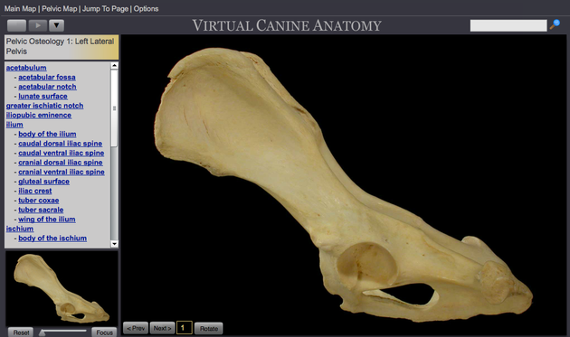
Here is another great resource to help you understand hip anatomy, from the vet school at Colorado State University. These are photographs of actual dissections, but if you stick to the photos of the skeletons you should be fine. After you click on this link, select the photos of "The Pelvic Limb" (the hind leg, middle photo of the three on the right).
http://www.cvmbs.colostate.edu/vetneuro/VCA3/vca.html
On the next screen, hover your mouse over the second photo from the left of the pelvic bone ("Osteology"), and a tree of the bones of the leg and pelvis will drop down. Select the first on from the top, the pelvis itself.
There is lots of really cool stuff you can do with this. First, on the lower left there is a slider that will let you zoom in and out on any area of the bone. You can click "next" and "previous" to go through a series of photographs of the pelvis from different angles. But the really fun one is the "Rotate" button. Click on that, and you will be able to rotate the pelvis in three dimensions as well as zoom in and out.
When the animal is standing, the top of the acetabular rim will be supporting the head of the femur as the weight-bearing surface. When you think about the weight of a dog, and the pounding the pelvis must endure during running and jumping, you can appreciate the magnitude of the forces the rim of the hip socket must endure.
You can rotate the pelvis so it is oriented just as it would be during the pelvic x-ray for a hip scan. When you do this, you will be able to clearly see the pair of hip sockets on either side (and you'll also see the acetabular notch). Now if you move the pelvis just a tiny bit, you can see how much it changes your view of those hip sockets. Even a tiny shift can make the socket on one side look much deeper and the one on the other side shallower. This is why getting the proper positioning of the dog during the hip x-ray is so critical.
If you click on the anatomical terms in the list on the left, you will see the particular bone identified on the skeleton. There is also some text that will appear in the box below the photograph which provides some information about the bone, what it does, and the various other bones, ligaments, and muscles that attach to it.
You can go back to the index and pick photo of the femur articulated with the socket to see how they fit together. As before, there are multiple views and you can zoom in and out as well as rotate.
This is a great resource for helping you to understand the anatomy of the hip joint, and you can explore other areas of interest as well - the elbow, shoulder, stifle, foot, skull...well, pretty much the whole dog! Enjoy!
You can learn lots more about hip and elbow structure and function in ICB's online course "Understanding Hip and Elbow Dysplasia".
The next class begins January 4, 2016.
Check out
ICB's online courses
*******************
Coming up NEXT -
Understanding Hip & Elbow Dysplasia
Next class starts
4 January 2016
Managing Genetics for the Future
Next class starts
18 January 2016
Sign up now!
***************************************
Visit our Facebook Groups
ICB Institute of Canine Biology
...the latest canine news and research
ICB Breeding for the Future
...the science of dog breeding


